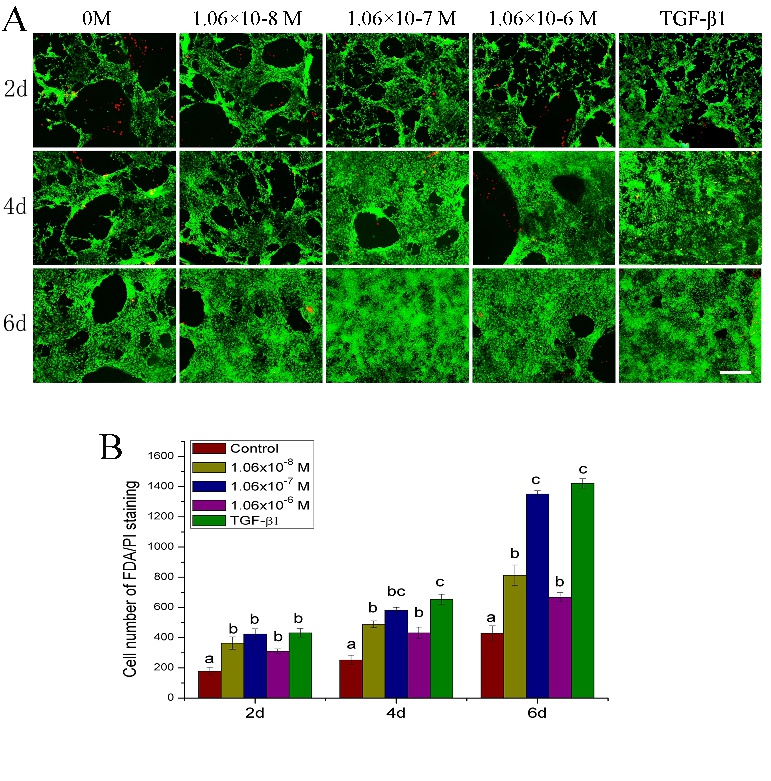
Fig. 1. (A) Confocal laser scanning microscopy images showing the viability of chondrocytes cultured in vitro alone (Control) or with ZXHA-C (1.06×10⁻⁸ M, 1.06×10- 7 M, 1.06×10⁻⁶ M) and TGF-β1 (T=15 ng/mL) for 2, 4 and 6 days. Cell seeding density: 2×10⁴/mL (original magnification ×40, scale bar was 100µm). (B) Cell number calculated and analyzed on the basis of FDA/PI staning images. Cell numbers were counted in one image by using the „cell calculating“ toolbar in the Nikon A1 software. The data represent the mean ± SD of three independent ulcutre experiments. Bars with different letters are significantly different from each other at P < 0.05.